Posts

Showing posts from July, 2014

#10.Tests for carbohydrates

Image
Tests for Reducing sugars, Non-reducing sugar and Starch . 1.  Reducing sugar ( Ben edict's test) All  monosaccharides  and most  disaccharides  (except sucrose) will reduce  blue   CuSO4 (II), producing a precipitate of  red   Cu 2 O (I). Benedict’s reagent is an aqueous solution of Cu SO4(II), Na 2 CO3 and sodium citrate.  2 cm³ test solution +  ≥  2 cm³ Benedict’s reagent.  Shake, and heat for a few minutes at 95°C in a water bath. The mass of precipitate or intensity of the colour indicates the amount of reducing sugar present  ---> the test is semi-quantitative.  2. Non-reducing sugar ( Benedict's test)  Principles: Sucrose  is a non-reducing sugar (not reduce  CuSO4) --->  Benedict's test (-) . If it is hydrolysed to form  glucose   and  fructose  ---> Benedict's test (+) .  So sucrose is the only sugar that will give a (-) Benedict's test before hydrolysis and a (+) test afterwards.  Steps:  Test a sample for reducing sugars to be sure it does not

#9. Carbohydrates - Polysaccharides

Image
Molecules contain hundreds/thousands of monosaccharides linked into long chains. Molecules are enormous --> the majority do not dissolve in water --> good for  storing energy  ( starch and glycogen ) or for  forming strong structures  ( cellulose ). 1. Storage Polysaccharides Glycogen  (in  animals and fungi) Made of α-glucose molecules linked together by  glycosidic bonds .  Most of the bonds are α1-4 links (C1 on one glucose + C4 on the next)  There are some 1-6 links, which form branches in the chain.  The bonds can be hydrolysed by carbohydrase enzymes to form monosaccharides, used in respiration.  The branches increase the rate of hydrolysis. 1-4 and 1-6 links in Glycogen. Starch (in plants) A mixture of  amylose  and  amylopectin . Both forms of starch are polymers of α- Glucose. Natural starches contain 10-20% amylose and 80-90% amylopectin. Amylose   molecule is a very long chain with 1-4 links. The chain coils up into a spiral, held in shape by H bonds between the gluc

#8. Carbohydrates - Monosaccharides, disaccharides

Image
Carbohydrates: - Sugar polymers - Molecules contain C, H, O atoms - H atoms are twice as many as C or O atoms  (C 6 H 12 O 6 ) Monosaccharides  The simplest carbohydrates  They are sugar: C = 3 = triose   C = 4 = tetrose  C = 5 = pentose C = 6 = hexose Examples of hexose sugars: glucose, fructose, galactose (C 6 H 12 0 6) Molecules often have the form of a ring, made up of some C atoms and one O atom.  Glucose molecules has 2 forms: α-glucose and β-glucose. Disaccharides  Different disaccharides can be formed by linking different monosaccharides. The bond that joins them together =  glycosidic bond.   Condensation reactions  (dehydration): 2  monosaccharides covalently joined; H 2 0 is formed. Hydrolysis reaction (splitting by water): disaccharides are split into 2 monosaccharides by breaking the glycosidic bond; a molecule of H 2 0 is added.  Functions of monosaccharides and disaccharides   Good sources of energy in living organisms, used in respiration for making ATP. Transportable

#7.2 Biological molecules - Syllabus 2016

2.1    Testing for biological molecules 2.2    Carbohydrates and lipids 2.3    Proteins and water This section introduces carbohydrates, proteins and lipids: organic molecules that  are important in cells. Nucleic acids  are covered in a separate section. Biological molecules are based on the versatile element carbon.  This section explains  how macromolecules, which have a great  diversity of function  in organisms, are assembled from smaller  organic molecules such  as glucose, amino  acids,  glycerol and fatty acids. Life as we know it would not be possible without water. Understanding the properties of this extraordinary molecule is an essential part of any study  of biological molecules. The emphasis in this section is on the relationship between molecular structures and their functions. Some of these ideas  are continued in other  sections, for example, the functions of haemoglobin in gas transport in Transport of mammals, phospholipids in membranes in Cell membranes and transpo

#7.1 Biological molecules - Syllabus 2015

• Structure of carbohydrates, lipids and proteins and their roles in living organisms • Water and living organisms Learning Outcomes Candidates should be able to: (a) [PA] carry out tests for reducing and non-reducing sugars (including using colour standards as a  semi-quantitative use of the Benedict’s test), the iodine in potassium iodide solution test for starch, the  emulsion test for lipids and the biuret test for proteins; (b) describe the ring forms of α-glucose and β-glucose (candidates should be familiar with the terms  monomer, polymer and macromolecule); (c) describe the formation and breakage of a glycosidic bond with reference both to polysaccharides and to  disaccharides including sucrose; (d) describe the molecular structure of polysaccharides including starch (amylose and amylopectin),  glycogen and cellulose and relate these structures to their functions in living organisms; (e) describe the molecular structure of a triglyceride and a phospholipid and relate these stru

#6 Summary of Cell structure

Image
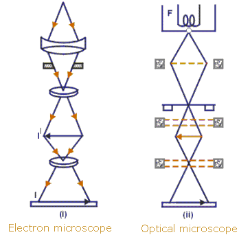
The basic unit of life, the cell, can be seen clearly only with the aid of microscopes. The light microscope uses light as a source of radiation, whereas the electron microscope uses electrons. The electron microscope has greater resolution (allows more detail to be seen) than the light microscope, because electrons have a shorter wavelength than light. With a light microscope, cells may be measured using an eyepiece graticule and a stage micrometer. Using the formula A= I/M, the actual size of an object (A) or its magnification (M) can be found if its observed (image) size (I) is measured and A or M, as appropriate, is known. All cells are surrounded by a partially permeable cell surface membrane that controls exchange between the cell and its environment. All cells contain genetic material in the form of DNA, and ribosomes for protein synthesis. The simplest cells are prokaryotic cells, which are thought to have evolved before, and given rise to the much more complex and much larger

#5. Plan diagrams of tissue and organ, prokaryotic and eukariotic cells

Image
An  organ  usually contains many different types of cells. These are arranged in a particular pattern characteristic of the organ, with cells of a similar type found together, forming distinctive  tissues. A plan diagram shows the distribution of  tissues  in an organ, not individual  cells . A cross section of leaf, stem and root.  Prokaryotes and Eukaryotes  There are only 2 basic types of cells, primitive prokaryotes and the more complex eukaryotes. Prokaryotic cells  ( Pro=“before”, karyon = “nucleus” ) are evolutionarily ancient. They were here first and for billions of years were the only form of life. Today most life is prokaryotic, and these cells are supremely successful. All bacteria and bacteria-like Archaea are prokaryotic organisms. Eukaryotes (Eu=“true”, karyon= “nucleus” ) can be single celled or multi-cellular organisms. Eukaryotic cells are more complex, having evolved from a prokaryote-like predecessor. Most of the living things that we are typically familiar with ar