Posts

Showing posts with the label Cell structure

#6 Summary of Cell structure

Image
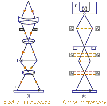
The basic unit of life, the cell, can be seen clearly only with the aid of microscopes. The light microscope uses light as a source of radiation, whereas the electron microscope uses electrons. The electron microscope has greater resolution (allows more detail to be seen) than the light microscope, because electrons have a shorter wavelength than light. With a light microscope, cells may be measured using an eyepiece graticule and a stage micrometer. Using the formula A= I/M, the actual size of an object (A) or its magnification (M) can be found if its observed (image) size (I) is measured and A or M, as appropriate, is known. All cells are surrounded by a partially permeable cell surface membrane that controls exchange between the cell and its environment. All cells contain genetic material in the form of DNA, and ribosomes for protein synthesis. The simplest cells are prokaryotic cells, which are thought to have evolved before, and given rise to the much more complex and much larger ...

#5. Plan diagrams of tissue and organ, prokaryotic and eukariotic cells

Image
An  organ  usually contains many different types of cells. These are arranged in a particular pattern characteristic of the organ, with cells of a similar type found together, forming distinctive  tissues. A plan diagram shows the distribution of  tissues  in an organ, not individual  cells . A cross section of leaf, stem and root.  Prokaryotes and Eukaryotes  There are only 2 basic types of cells, primitive prokaryotes and the more complex eukaryotes. Prokaryotic cells  ( Pro=“before”, karyon = “nucleus” ) are evolutionarily ancient. They were here first and for billions of years were the only form of life. Today most life is prokaryotic, and these cells are supremely successful. All bacteria and bacteria-like Archaea are prokaryotic organisms. Eukaryotes (Eu=“true”, karyon= “nucleus” ) can be single celled or multi-cellular organisms. Eukaryotic cells are more complex, having evolved from a prokaryote-like predecessor. Most of the liv...

#4. Cell structure and function

Image
Describe and interpret drawings and photographs of typical animal and plant cells. Note that plant cells are always surrounded by a cell wall made of cellulose,  never found around animal cells.  Typical animal and plant cells as seen using an electron microscope: 1. Functions of membrane systems and organelles The plasma membrane ( cell surface membrane ) controls what enters and leaves the cell. Many membranes within the cell help to make different compartments for different chemical reactions to take place. The nucleus is surrounded by  nuclear envelope (pair of membranes).  The nucleus contains chromosomes , with very long molecule of DNA (DNA determines the sequences of amino acids to form protein molecules).  A darker area in the nucleus (no membrane) is called nucleolus : here new ribosomes are made, following a code on part of the DNA. Ribosomes (made of RNA & protein) are found free in the cytoplasm + attached to rough endoplasmic reticulum (R...

#3. Microscopy

Image
Most cells are very small, and their structures can only be seen by using a microscope . 1. Light microscopes light rays pass through the specimen on a slide  focused by an objective lens and an eyepiece lens .  ---> magnified image of the specimen on the retina of your eye/screen/camera . 2. Electron microscopes uses beams of electrons   specimen very thin,  placed in a vacuum  to allow electrons to pass through it.  electrons are focused onto a screen/photographic film ---> magnified image of the specimen. 3. Magnification and Resolution Amount of magnification depends on the resolution of the microscope (ability to distinguish 2 objects as separate).  The smaller the objects that can be distinguished --> the higher the resolution.  wavelength: beam of electrons  <<<  light          resolution:   electron microscope >>> light microscope with electron microscope,...